English
 German
 Chinese
Technology for super-resolution microscopy
Optical microscopy methods which break the diffraction limit by Ernst Abbe are maned as so-called super-resolution techniques. Unlike the classical light microscopy which has a resolution limit in the range of several hundred nanometers, super-resolution microscopy offers the opportunity to resolve details with a precision of a few nanometers without being invasive like electron microscopy or atomic force microscopy. Therefore these techniques became a more and more important tool in the cell imaging.
Common techniques are e.g. SIM (structured illumination microscopy), STED (stimulated emission depletion) or localization based methods. Whereas SIM and STED generate the higher resolution by applying technical tricks on the hardware side, localization based techniques work by controlling the fluorescent signal of the used probes enabling a temporal separated detection. This can be done either photophysical as in PALM and (d)STORM for instance or on the base of binding kinetics like in DNA-PAINT. By a clever combination of the hardware- and the probe-based ansatz the technique MINFLUX (maximally informative luminescence excitation probing) gains an even higher resolution. Moreover there is a huge number of further resolution improving techniques applied in the field of biological imaging.
However, due to the lack of standardized methods for measuring and quantifying the resolution obtained by such a system, the user could not be sure that the used microscope provides the resolution which is required for his experiment.
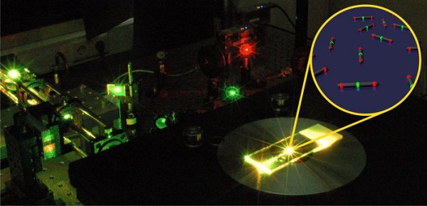
Nanorulers for super-resolution systems
Our innovative nanorulers now allow a fast, easy and precise determination of resolution values in any common super-resolution system (STED, SIM, (d)STORM, DNA-PAINT) as well as in diffraction limited applications (GATTA-Confocal). On the one hand the instantaneous visible, significant result makes it easy for users to proof their microscope’s resolution and on the other hand provides an opportunity for microscope manufacturers to demonstrate the performance of their products in an eye-catching way.
Fluorescent beads for optical microscopy
Additionally, GATTAquant developed point-like light sources performing the highest brightness density in the world. These gattabeads are ideal tools for exact and quantitative characterizations of super-resolution microscopes (especially STED systems) or as reference points for drift correction or particle tracking. Hereby gattabeads are not only much brighter than conventional beads of comparable size but also provide a higher homogeneity and flexibility.
Custom Labels
Fluorescence detection is used in many different areas. The requirements on the label are as diverse as the applications themselves. Essential parameters are, for example, the brightness, color, stability and size of the label. However, specific requirements such as ambient and buffer conditions, compatibility with a specific biological or chemical process can also play a decisive role in the selection and design of a label.
At GATTAquant, we bring with us broad expertise in physics, chemistry and biology to develop individual solutions together with our customers.
© 2026 GATTAquant GmbH
contact
GATTAquant GmbH
Staffelseestraße 8
DE-81477 München
Phone: +49 89 2153 720 80
INFO@GATTAQUANT.COM
funded by

Payment Options